Femoroacetabular impingement (FAI) is a condition in which extra bone grows along one or both of the bones that form the hip joint — giving the bones an irregular shape. Because they do not fit together perfectly, the bones rub against each other during movement. Over time this friction can damage the joint, causing pain and limiting activity.
What is the anatomy of the hip?
The hip is one of the body's largest joints. It is a "ball-and-socket" joint. The socket is formed by the acetabulum, which is a part of the large pelvis bone. The ball is the femoral head, which is the upper end of the femur (thighbone).
The bone surfaces of the ball and socket are covered with articular cartilage, a smooth, slippery substance that protects and cushions the bones and enables them to move easily.
What are different types of femoroacetabular impingement (FAI)?
There are three types of femoroacetabular impingement: pincer, cam, and combined impingement:

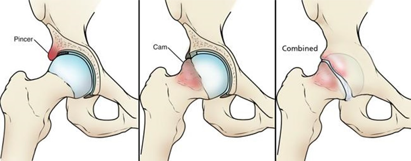
(Left) Pincer impingement. (Center) Cam impingement. (Right) Combined impingement.
- Pincer - This type of impingement occurs because extra bone extends out over the normal rim of the acetabulum. The labrum can be crushed under the prominent rim of the acetabulum.
- Cam - In cam impingement the femoral head is not round and cannot rotate smoothly inside the acetabulum. A bump forms on the edge of the femoral head that grinds the cartilage inside the acetabulum.
- Combined - Combined impingement just means that both the pincer and cam types are present.
What causes femoroacetabular impingement?
FAI occurs because the hip bones do not form normally during the childhood growing years. It is the deformity of a cam bone spur, pincer bone spur, or both, that leads to joint damage and pain. When the hip bones are shaped abnormally, there is little that can be done to prevent FAI. Some people may live long, active lives with FAI and never have problems. When symptoms develop, however, it usually indicates that there is damage to the cartilage or labrum and the disease is likely to progress.
Because athletic people may work the hip joint more vigorously, they may begin to experience pain earlier than those who are less active. However, exercise does not cause FAI.
What are symptoms of femoroacetabular impingement?
The most common symptoms of FAI include:
- Pain
- Stiffness
- Limping
Pain often occurs in the groin area, although it may occur toward the outside of the hip. Turning, twisting, and squatting may cause a sharp pain. Sometimes, the pain is just a dull ache.
How is femoroacetabular impingement diagnosed?
Impingement Test
As part of the physical examination, your doctor will likely conduct the impingement test.
For this test, your doctor will bring your knee up towards your chest and then rotate it inward towards your opposite shoulder. If this recreates your hip pain, the test result is positive for impingement.
Imaging Tests
Your doctor may order imaging tests to help determine whether you have FAI.
X-rays. These provide good images of bone, and will show whether your hip has abnormally shaped bones of FAI. X-rays can also show signs of arthritis.
Computed tomography (CT) scans. More detailed than a plain x-ray, CT scans help your doctor see the exact abnormal shape of your hip.
Magnetic resonance imaging (MRI) scans. These studies can create better images of soft tissue. They will help your doctor find damage to the labrum and articular cartilage. Injecting dye into the joint during the MRI may make the damage show up more clearly.
Local anesthetic. Your doctor may also inject a numbing medicine into the hip joint as a diagnostic test. If the numbing medicine provides temporary pain relief, it confirms that FAI is the problem.
How is femoracetabular impingement treated without surgery?
- Activity changes. Your doctor may first recommend simply changing your daily routine and avoiding activities that cause symptoms.
- Non-steroidal anti-inflammatory medications. Drugs like ibuprofen can be provided in a prescription-strength form to help reduce pain and inflammation.
- Physical therapy. Specific exercises can improve the range of motion in your hip and strengthen the muscles that support the joint. This can relieve some stress on the injured labrum or cartilage.
Departments and Programs Who Treat This Condition
Orthopedic Surgery
Hip and Knee Arthroplasty (Replacement)
Musculoskeletal Medicine and Pain Management Program
Hip Preservation
 en
en 
 Français
 Deutsch
 Italiano
 Español
 Tiếng Việt
 Kreyol ayisyen